This was a prototype of a transcutaneous (through-the-skin) sensor for blood glucose and blood alcohol.
Optical and optomechanical design, detection and control electronics and software, prototype construction: first spectra were taken 5/16/2013, and technology transfer to a contract engineering firm was essentially completed 6/14/2013.
Most notable was the schedule requirement: from a standing start, in less than 6 weeks’ work, I did a complete photon budget, designed and built all of the optics and electronics, wrote all the software, integrated and shipped the system. It worked great.
A bit of a departure from our usual fare: a low cost, high speed sensor for detecting blood spots in eggs using spectral differencing. I’m going to be doing the firmware as well as the optics and electronics, and this will be the first actual client work for our newly qualified PCB designer, Magdalen.
Competing devices use xenon flashtubes and very expensive photodiodes, but still need a lot of calibration and tweaking. That makes it a good candidate for our signature technique: A really careful photon budget followed by a design that actually reaches the theoretical optimum performance.
This is a research project with a small division of a very large manufacturer. It’s a complete optical/electronic design to measure optical absorbance in a rapidly moving assay plate.
This is a smaller job for a sensor manufacturer, interesting mostly for the size, weight, and power (SWaP) requirements. It has to be shot noise limited above 500 pA with a 1600 pF photodiode. (This is possible only because the bandwidth is relatively small.)
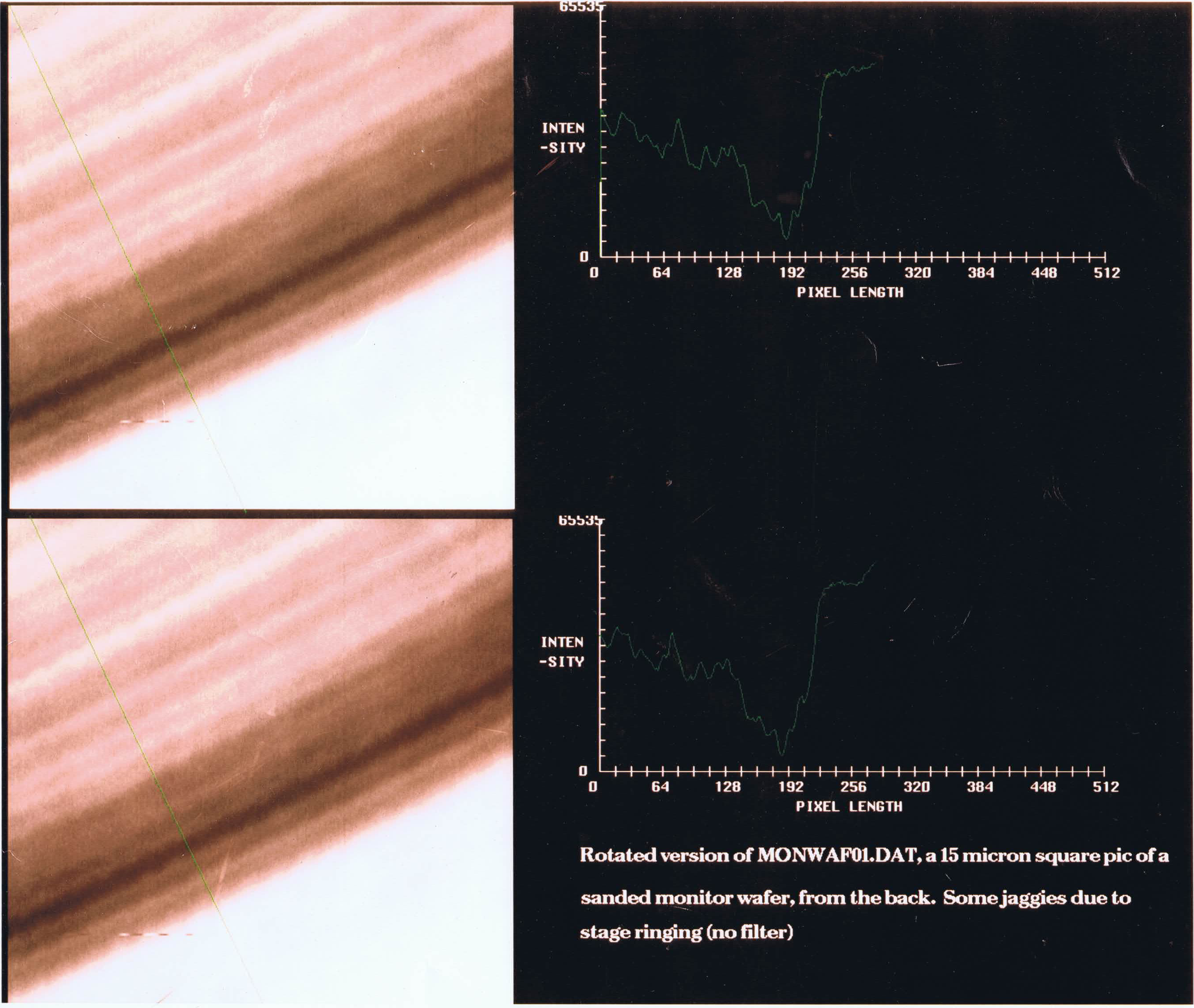
This is a very exciting development, in cooperation with a semiconductor equipment manufacturer. Back in 1989, my colleagues and I started working on a contact-lens microscope for looking into the interior of silicon chips through the back surface. In 1992 I took a picture (shown above) at a numerical aperture of 2.5, with resolution equivalent to NA 3.5 due to a confocal design.